Application Examples
Organoid Pre-processing
High-quality single-cell suspension is critical for organoid culture success. The MicroJet Dissociator provides standardized solutions with gentle dissociation, efficient enrichment, and precise control.
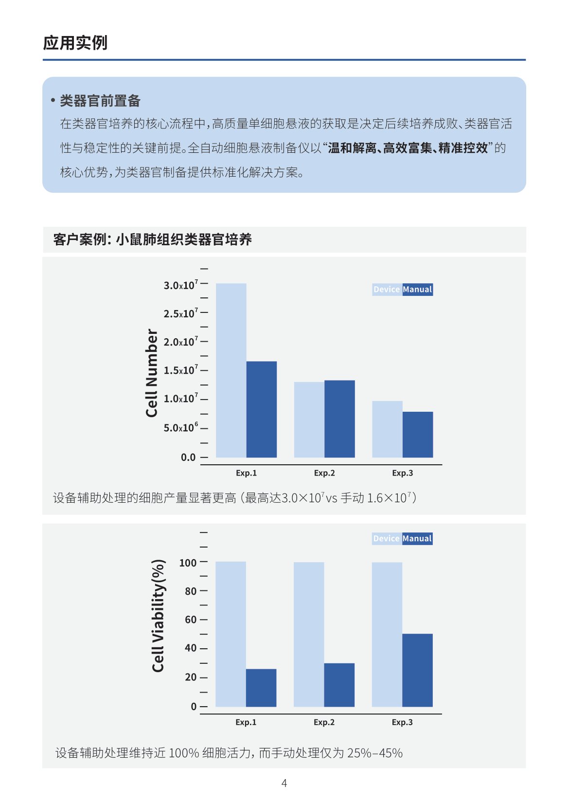
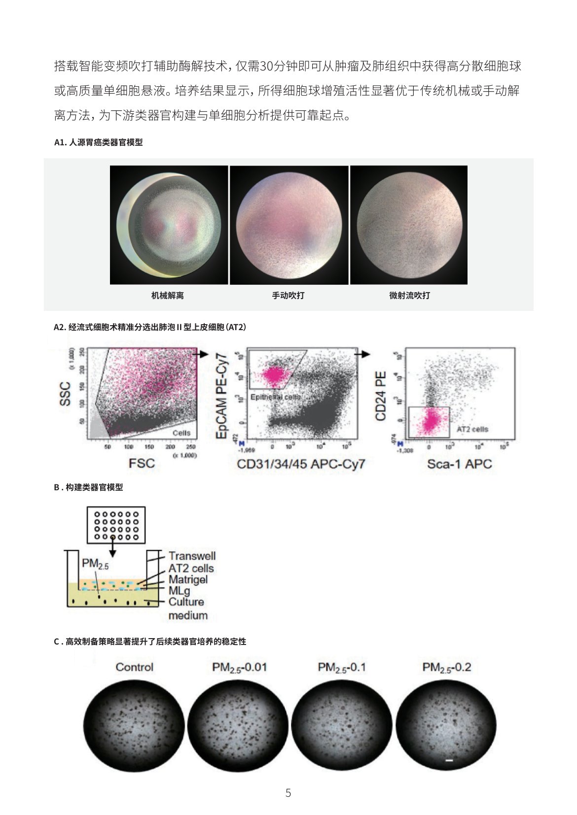
Near 100% cell viability with instrument-assisted processing (vs. 25–45% manual). Cell yields up to 3.0×10⁷ (vs. 1.6×10⁷ manual). FACS-sorted AT2 cells for organoid models with significantly enhanced culture stability.
Enhanced Cell Staining
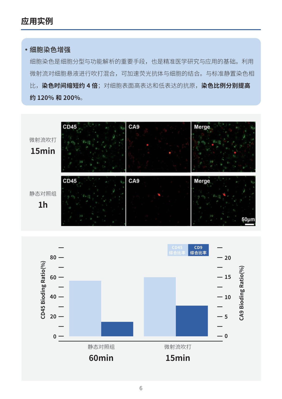
MicroJet mixing accelerates antibody binding — 4× faster staining vs. static incubation. Staining ratios improved ~120% and ~200% for low and high expression antigens respectively.
Single-Cell Analysis
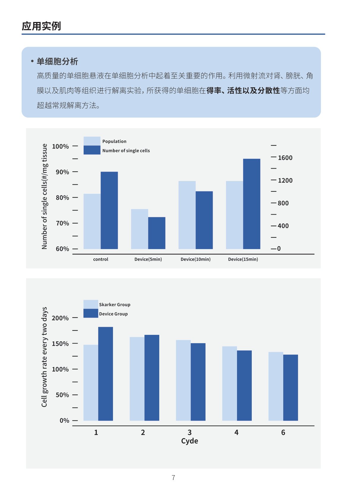
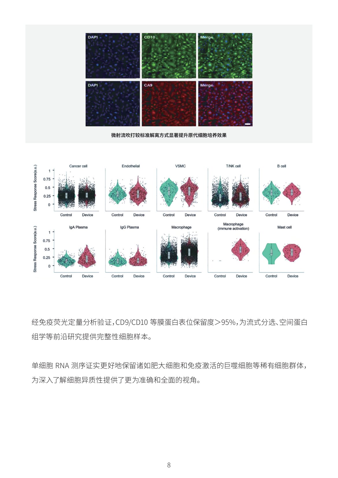
Superior yield, viability, and dispersion vs. conventional methods for kidney, bladder, cornea, and muscle tissues. >95% membrane protein epitope retention (CD9/CD10) for flow sorting and spatial proteomics.
Primary Cell Culture
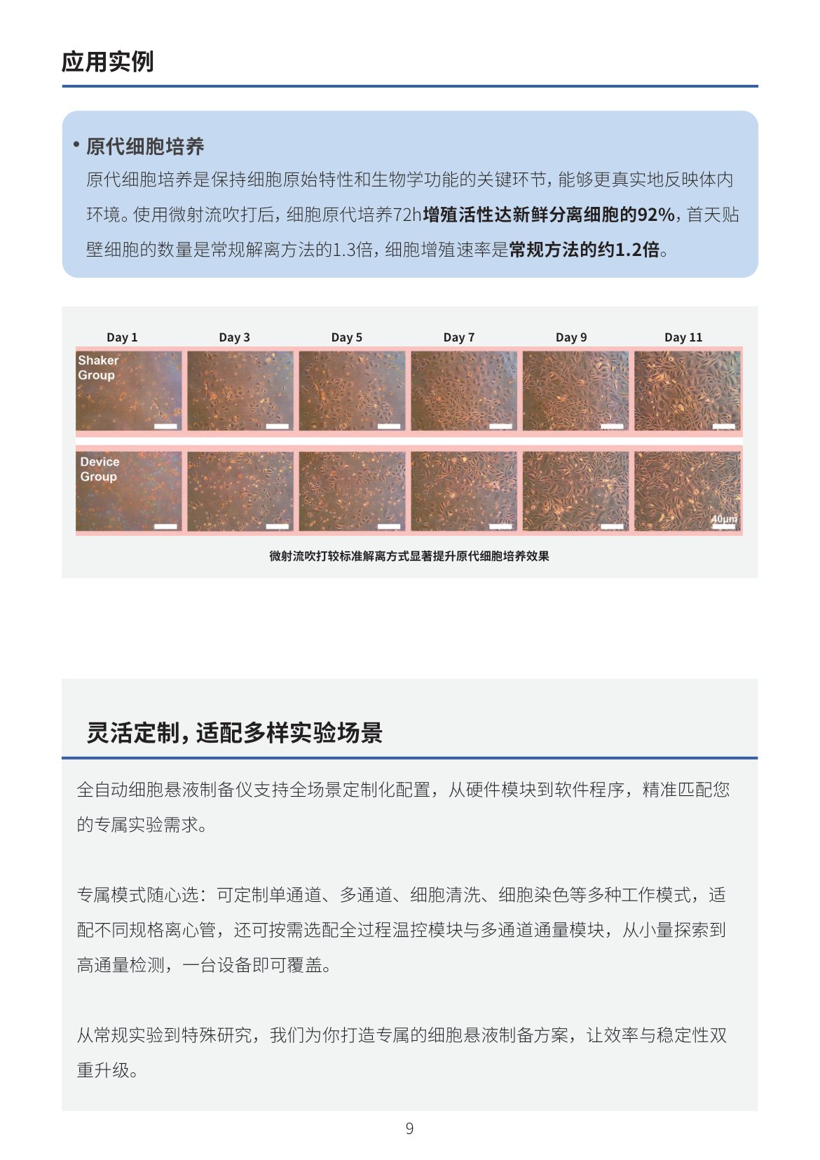
72h proliferation at 92% of fresh cells, 1.3× day-1 attachment, 1.2× proliferation rate vs. conventional methods.
Flexible Customization
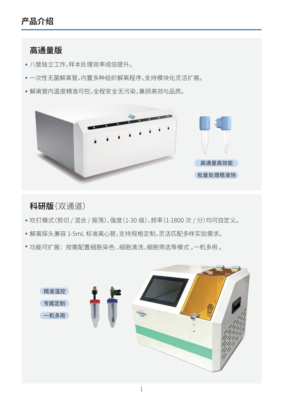
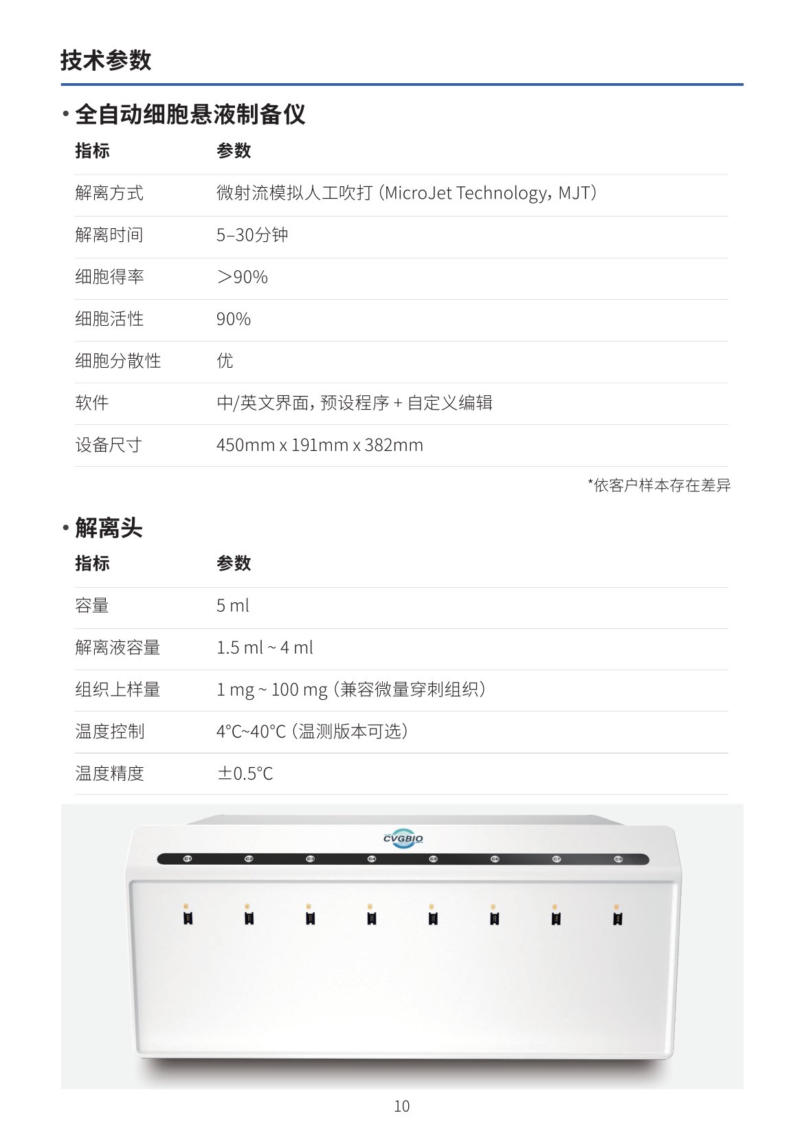
Full-scenario customization — single/multi-channel, washing, staining modes for various tube formats. Optional temperature control and multi-channel throughput modules. From exploratory research to high-throughput screening, one instrument covers all needs.
Partner Institutions
Harvard Medical School · Yale University · Tsinghua University · UMass · Max Planck Institute · University of Twente · University of Rome